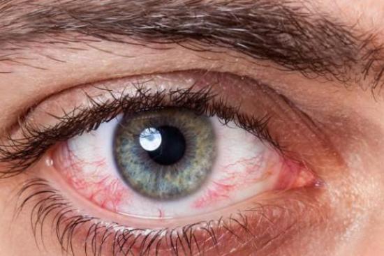
Thanks to many innovative treatments that prevent blindness and eye diseases, vision loss is about to become history. One of the most common diseases that lead to visual impairment is Retinitis Pigmentosa (RP). It is a genetic mutation that causes a dysfunction of the retinal cells, those that capture light. Hence, retinitis pigmentosa treatment is necessary to correct the visual disorders and subsequently regain clear vision.
- People with poor night vision
- People with a family history (mutation of the gene coding for retinal proteins)
- People with peripheral vision
- Allergic reactions
- Eye inflammation
Retinitis Pigmentosa treatment cost in Istanbul, Turkey
For patients who have become blind due to retinitis pigmentosa, the bionic eye represents an effective solution that aims to improve the quality of their life. With our ophthalmology clinics, the patient could regain autonomy by visualizing obstacles and objects.
Added to that, the new CRISPR technology can be used in future retinitis pigmentosa treatment. The technique can repair the genetic mutation that is the main factor of the disease. It consists of replacing the mutated gene with a healthy one to restore the function of the proteins translating the genetic code. It has great potential, but it is not 100% accurate. Furthermore, it must be verified that stem cells differentiate correctly into retinal cells. Besides, it is also necessary to make sure that CRISPR does not cause undesired genetic changes in the cells. We assure you the cheapest costto carry out your intervention
Best Clinics with Verified Reviews
- Network of 25 Clinics in Ophthalmology
- 2 Clinics in Germany and 1 in Amsterdam
- 96,000 patients in 2017
- Grand Hospital founded in 1999
- 200 Beds
- 53,000 square meters
- Member of the American Hospital Association (AHA)
- Capacity of 230 beds
- The best hospital in Ankara
What is retinitis pigmentosa?
Retinitis Pigmentosa (RP) is the photoreceptors' (cones and rods) degeneration. It is also known as pigmentary retinopathy, tapetoretinal degeneration, and rod-cone dystrophy. The patient, who suffers from rod deficiency, experiences visual impairment in the dark, especially when moving from luminous to dark environments. A simple eye exam, in our clinics in Turkey, would help to detect the presence of degenerative cells in the retina at the best costs.
What Causes rod-cone dystrophy
Several factors may lead to the onset and development of pigmentary retinopathy. The main causes of retinitis pigmentosa (rod-cone dystrophy) can be:
Heredity causes
Retinitis pigmentosa is a genetic condition that affects both eyes. Transmission can be transmitted in about 50% of patients with predominantly autosomal recessive or dominant transmission. About 10% of patients have a transmission on the X chromosome. If it occurs in one eye, the other eye usually experiences the same condition over the years. The genetic factor seems to be the main one, and retinitis pigmentosa is hereditary in most cases.
Congenital disease
Retinitis pigmentosa is usually diagnosed during adolescence, but may also be present at birth. The second congenital type is usually quite stable and not progressive. It is a group of inherited retinal disorders characterized by progressive degeneration of the light-sensitive cells (photoreceptors) in the retina, leading to visual impairment and, in severe cases, blindness. Thus, this condition is often associated with more severe vision loss compared to other forms of RP.
Systemic diseases
Retinitis pigmentosa is sometimes associated with other systemic diseases, such as Usher syndrome, Bardet-Biedl syndrome, Refsum disease, and Mitochondrial disorders. It's important to note that these systemic diseases are relatively rare and may not be present in all individuals with RP. The specific association between RP and these systemic conditions can vary, and additional symptoms and complications may be present. Genetic testing and a thorough medical evaluation by a healthcare professional specializing in genetic disorders or ophthalmology are necessary for an accurate diagnosis and appropriate management.
Complications of Pigmentary Retinopathy
Retinitis Pigmentosa in some patients may have certain complications, such as:
- Cataract: cataract removal can be performed for patients who are good candidates for the implantation of an intraocular lens;
- Blindness: vision loss is due to the failure of the retinal function.
Symptoms of Pigmentary Retinopathy
There are many symptoms associated with Retinitis pigmentosa. The first symptom of the reduced function of the photoreceptors with the difficulty of adaptation to the dark (night blindness). The patient experience this symptom when transitioning from a more luminous environment to one less luminous. This is often associated with a reduction in the peripheral field of view that causes it to bump against obstacles laterally. Patients find it difficult to identify the people placed on the side. In a later stage, dazzle may appear and a reduction of the sight that can even reach blindness can arise.
Other symptoms associated with RP
People with RP may experience other health problems that arise at a certain stage throughout the disease development:
- Hard time reading printed text;
- Night vision impairment;
- Tunnel vision, central vision, or peripheral vision;
- Difficulty recognizing shapes in images;
- Glare in the eyes.
How is Retinitis Pigmentosa Diagnosed?
If you suspect that your visual difficulties are due to a case of retinitis pigmentosa, you should contact the ophthalmologist. Only the specialist can carry out all the necessary examinations to accurately identify the pathology.
Fundoscopic exam
Its purpose is to evaluate the retinal morphology and to investigate the presence of characteristic pigment spots on the retinal surface. Some forms of retinitis do not include the presence of spots on the bottom of the eye as symptoms. The ophthalmologist usually associates the diagnosis with specific instrumental examinations that aim to evaluate the rods' function. It is also associated with electroretinography (ERG) which records retinal electrical activity.
Visual field test
The best eye doctors can evaluate the retinal sensitivity to a luminous stimulus in the various areas of the retina. It is useful to have objective documentation of the difficulties perceived by the patient. The visual field test evaluates the extent of visual capacity in space. On the other hand, optical coherence tomography OCT highlights the individual layers of the retina. Thus, the deficiencies of the rods can be highlighted.
Retinitis Pigmentosa Cure & Treatment options
Nowadays, there are several Retinitis Pigmentosa cure options available. Numerous therapeutic attempts have been made to treat retinitis pigmentosa, including medical means.
The retinitis pigmentosa therapy techniques include:
- Administration of vasodilators;
- Anticoagulants;
- Hyperbaric oxygen therapy;
- Ozone therapy;
- Vitamins and minerals;
- Hormones;
- Acupuncture;
- Ultrasound and electrical stimulation.
Options of Retinitis Pigmentosa Therapy: Which one is effective?
All the techniques aim at improving blood supply and retinal metabolic process, but none were effective. Some options can slow vision loss and even restore sight to some extent. The cost of the therapy varies depending on the method of the retinitis pigmentosa cure employed.
Best Methods
Some leading clinics and hospitals started using innovative ways in curing Retinitis Pigmentosa. These approaches include:
- Gene therapy;
- Anti-apoptotic agents;
- Ribozyme therapy.
Share this page
It is a genetic, chronic and progressive eye disease. It causes gradual degeneration of the photoreceptor cells in the retina, that are responsible for night vision (the rods).
- Vision problems when the light decreases (night blindness);
- Decreased visual field (tunnel vision);
- Color perception disorders (achromatopsia);
- In the most advanced cases, the disease can lead to cataracts (opacification of the crystallin).
- Wearing suitable protective and filtering glasses (anti UV);
- A supply of vitamin A and E to slow down the deterioration of cones and rods.
The epiretinal prosthesis in our partner clinics in Turkey is an "artificial retina" implanted in the patient's eye. Its role is to replace the photoreceptor cells of the retina (rods and cones).
The prosthesis is associated with small cameras (in the form of glasses) and a microcomputer placed in the patient's pocket. This device converts the visual information captured by the glasses into electrical signals, which are transmitted by radio waves to a receiver placed on the eye.