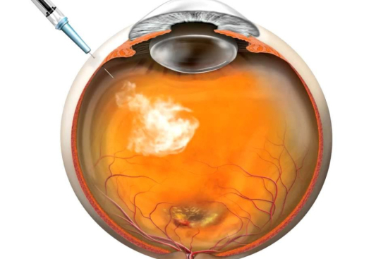


The retina separates from its supporting tissue. Vision blurs, sometimes disappears entirely.
This condition, known as retinal detachment, demands urgent surgical intervention to prevent permanent blindness. Without prompt repair, the light-sensitive cells begin to deteriorate irreversibly, typically within days or weeks.
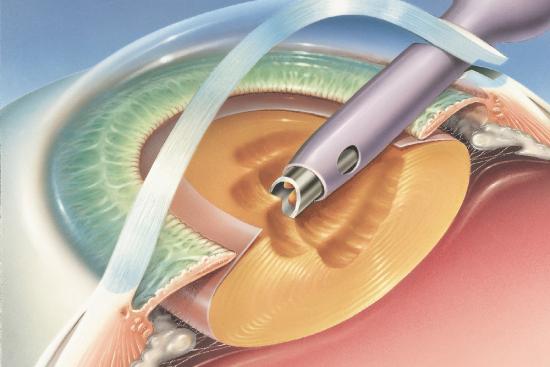
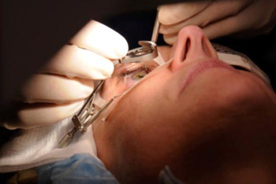
In Turkey, ophthalmologists perform specialized treatment using three main surgical approaches: vitrectomy (removing the gel inside the eye),pneumatic retinopexy (injecting an expanding gas bubble),and scleral buckle (placing a silicone band around the eye). Each technique addresses different detachment types and severity levels.
Patients report gradual vision improvement over months, not immediately after surgery (this matters psychologically). Recovery requires patience, strict eye positioning, and regular follow-up appointments.
Success rates hover around 87% for primary repairs, though some cases require additional procedures. Myopic patients, those with previous eye trauma, or individuals with diabetes-related complications face higher detachment risk and benefit from early consultation with a retinal specialist.
Retinal Detachment Surgery Cost in Turkey
Pricing varies based on surgical complexity and chosen technique. Turkish clinics offer transparent cost structures significantly lower than Western Europe or North America, without compromising quality or safety standards.
Scleral buckle with cerclage typically ranges from $2,800 to $3,500. This procedure suits straightforward detachments with single tears and involves less operative time than vitrectomy.
Vitrectomy costs between $3,800 and $5,200 depending on complexity, additional procedures needed, and whether gas or silicone oil tamponade is used. Complex cases with multiple tears or macula involvement fall toward the higher end.
Pneumatic retinopexy ranges from $2,200 to $3,200, making it the most economical option for eligible candidates. However, strict patient selection criteria limit its applicability.
These prices include surgeon fees, facility costs, anesthesia, and immediate post-operative care. Travel packages combining surgery with accommodation can reduce overall expenses. Many Turkish clinics offer payment plans or work with international insurance providers. Always request itemized quotes and verify JCI accreditation before committing.
Risks and Side Effects
Temporary eye and facial swelling, mild to moderate discomfort, and transient vision disturbances are common. Rare complications include infection, elevated eye pressure, or incomplete reattachment requiring revision surgery.
- Swelling and bruising around the eye (resolves within 2-3 weeks)
- Mild pain or foreign body sensation
- Temporary blurred or distorted vision
- Risk of recurrent detachment in 5-10% of cases


 LIV Vadistanbul")