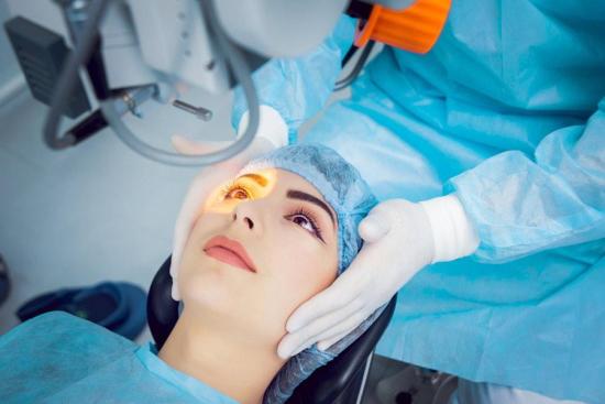
Corneal replacement is a surgical operation in which the diseased cornea will be changed by an artificial cornea. Keratoprosthesis is indicated after the failure of one or more corneal transplants from a donor.
Recently, a less invasive and non-invasive artificial cornea has been augmented, which can be employed in routine corneal blindness. Thus, the ophthalmic surgeon uses an artificial cornea to replace the defective one in place of using donor tissue for transplantation (conventional corneal transplant). This operation is performed to correct the patients' vision whose corneas are severely damaged due to inborn malformations, or following trauma affecting the outer layer of the eye.
Contact us and get a free quote for your keratoprosthesis in Turkey in one of our partner clinics!
Patients with:
- Vision less than 20/200 in the affected eye
- A failed corneal transplant using a corneal donor and who have little vision or who do not have vision
- Birth defects, non-autoimmune diseases, and other eye problems
- Glaucoma and implant extrusion
- Sudden inflammation of the vitreous can cause severe reduction in vision
- Inflammation can occur in the tissues of the eye
- Corneal transplant
- One to two hours
- 98%
Eye surgery course in Istanbul, Turkey: Price & clinics
The patient will be oriented to one of our ophthalmic partner clinics or hospitals or a laser center to undergo his eye operation. After a physical examination, he will be taken to the operating room. Keratoprosthesis is performed under general or local anesthesia in Turkey at an affordable price.
A blepharoscope is used to keep the eye open throughout the surgery. Indeed, certain lubricants can be used to prevent dry eyes. Depending on the type of keratoplasty used, the surgery may involve the replacement of a full-thickness cornea or the placement of an intra-lamellar implant. Using the Alphacor method, a manual incision is worn to design a corneal pocket and an awl to create an opening through the posterior cornea. The Alphacor is inserted into the corneal sinus to allow biointegration after many months, a second procedure is assimilated to remove part of the anterior cornea and facilitate access of light to the retina. In the case of KeraKlear, the pocket is created after several months. A second procedure is used to remove part of the anterior cornea to allow light to reach the retina.
Usually, there will be a follow-up session a few days after the surgery, during which the patient's assessment is treated and adjustments will be made.
Best Clinics with Verified Reviews
- Network of 25 Clinics in Ophthalmology
- 2 Clinics in Germany and 1 in Amsterdam
- 96,000 patients in 2017
- Grand Hospital founded in 1999
- 200 Beds
- 53,000 square meters
Keratoprosthesis: definition of the procedure
Keratoprosthesis are constructed of invisible plastic by texture with optical characteristics. They vary in design, size, and even implantation methods may vary from one treatment center to another. This procedure is performed by ophthalmologists, often on an outpatient basis, thanks to the corneal transplantation practices evolution.
Guillaume Pellet de Koenigse, a French ophthalmologist, originally proposed the notion of an artificial cornea in 1789.
The different types of Keratoprosthesis
Many keratoprosthesis have been developed, but four models have currently been marketed: Boston keratoplasty, Osteotomy-Odonto-keratoplasty (OOKP),AlphaCor, and KeraKlear prosthetic keratoplasty.
Usage factors of the implantation
A corneal transplant is performed when the corneal pathology is not accessible to conservative treatments (rigid lenses, lasers, intracorneal rings) severely reduces visual sensitivity or is the cause of major ailments.
It is based on the location of the cornea to its entire extent, this technique is called transfixing cornea transplant or only partially (lamellar cornea transplant) it depends on the pathology.
The reasons for reduced visual acuity of corneal origin can be a loss of transparency (corneal edema, post-traumatic or infectious scar, for example, herpetic or following an infection under contact lenses.),or even an anomaly of irregular shape.
The indications for ophthalmic surgery
Indications for penetration of artificial corneas include:
- Treatment of patients with vision less than 20/200 in the affected eye.
- Patients with unsuccessful corneal transplants using a corneal donor and who have little vision or who do not have a vision.
- Patients who do not have access to the transplanted corneal tissue.
- Indications for non-penetrating corneal replacement include:
- Keratoconus
- Corneal atrophy
- Corneal scars unrelated to inflammation of the eye
- Corneal edema
- Loss of limbal stem cells
- Eclipse of corneal transplants with a non-inflammatory diagnosis (eg, keratoconus)
The preoperative examination
As before any surgery, the patient visits the ophthalmologist for a visual diagnosis and other tests. During the consultation, the ophthalmologist will examine the condition of the eye. Thus, it will also record the patient's medical history and other previous eye care. Therefore, the doctor will discuss the complications and the benefits of Keratoprosthesis surgery. Once the patient opts for surgery ophthalmic, the practitioner will ask them to also perform medical tests and laboratory tests, such as an x-ray, a slit lamp test, an EKG, an ultrasound B-scan. Or an A-scan. The date and time of the intervention will also be determined.
The conditions of the Keratoprosthesis procedure
Keratoprosthesis is indicated in certain conditions:
- Visibility less than 20/200
- The retina must be in place and there must be no severe suction of the optic nerve.
- Case of advanced glaucoma
The risks of ophthalmic surgery
Although the success rate of corneal replacement surgery is high, in very rare cases significant complications can occur:
- Glaucoma and the extrusion of implants
- Sudden inflammation of the vitreous can cause severe reduction in vision. However, this condition can be treated with antibiotics or with simple laser surgery.
- Inflammation can occur in the tissues of the eye. This condition is also can be treated.
Share this page